A Japanese research team has identified how iron toxicity and BRCA1 mutations contribute to the formation of precancerous giant-nucleus cells in the kidneys.
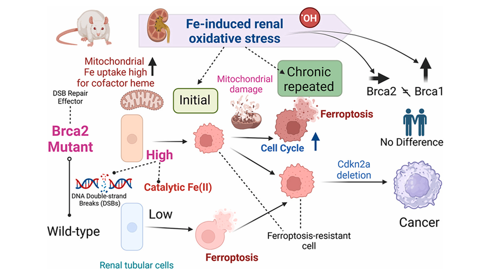
Iron is an essential micronutrient, but excess iron can damage cells and increase cancer risk. While many cells die from iron-induced oxidative stress, a subset survives and may become cancerous. Previously, the identity of these surviving cells was unclear.
A Japanese research team has demonstrated that giant-nucleus cells that survive iron-induced damage form the foundation of early-stage cancer. The team anticipates that these giant-nucleus cells could serve as early indicators of cancer, as they are easy to identify under a microscope. The study was published in the journal Redox Biology.
“Since the 1980s, scientists have spotted cells with abnormally large nuclei in the early phases of cancer triggered by oxidative stress, but their role in cancer development was unclear,” said Shinya Toyokuni, lead author and professor emeritus at Nagoya University Graduate School of Medicine.
To investigate the role of these giant-nucleus cells, Dr. Toyokuni, Assistant Professor Yingyi Kong, and their colleagues conducted experiments using both wild-type rats and rats carrying a single mutated BRCA1 gene.
Mapping iron-induced damage
Researchers injected ferric nitrilotriacetate, a carcinogenic iron compound, into rats to induce kidney damage. They then collected kidney tissue samples before the injection and one and three weeks afterward.
For the initial analysis, the team performed an in-depth examination of representative kidney tissue samples across the different time points. They subsequently validated their findings by analyzing additional animals and comparing the results with human cancer datasets.
The researchers used spatial transcriptomics to map gene activity in individual cells within tissue sections while preserving the tissue’s original structure. They also conducted computational analyses to quantify the size, shape, and density of cell nuclei in the same samples.
How surviving cells become cancerous
Within one week of iron administration, giant-nucleus cells emerged. These cells showed increased expression of cancer-related genes such as Myc and Met, and developed resistance to ferroptosis, an iron-dependent form of cell death.
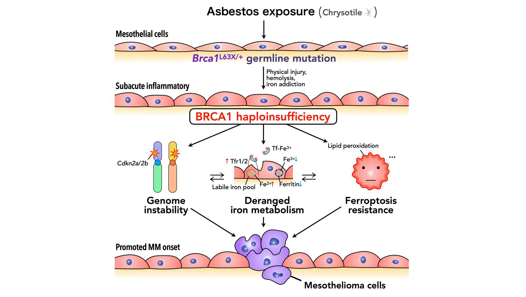
BRCA1-mutant rats developed giant-nucleus cells with distinct biological profiles from wild-type rats. This suggests that BRCA1 deficiency impairs DNA repair, allowing more precancerous giant-nucleus cells to survive.
Notably, even morphologically normal cells near giant-nucleus cells exhibited gene expression changes that may promote their survival. This suggests that large nuclei in kidney cells could signal early cancer development in surrounding tissue.
Additionally, kidney tissue from BRCA1-mutant rats showed a substantial increase in stromal cells (non-cancerous supportive cells, including immune and connective tissue elements) around damaged regions. This indicates a more pronounced alteration of the local tissue environment in these cases.
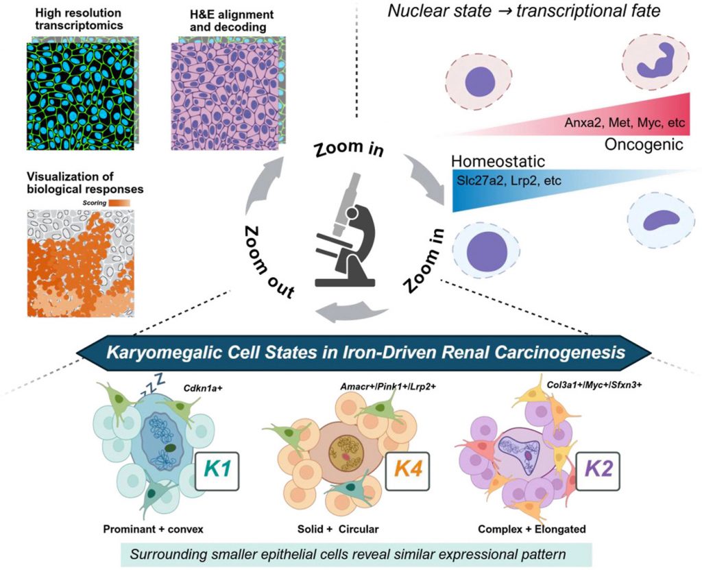
By analyzing gene activity and nuclear morphology, the researchers classified giant-nucleus cells into six types. One type had extremely large nuclei and signs of stress-induced growth arrest, consistent with profiles from previous studies. Another type, more common in BRCA1-mutant kidneys, showed increased cancer-related gene activity and elongated nuclei, indicating a potential precancerous state.
Additional electron microscopy and cellular respiration measurements confirmed that cells from BRCA1-mutant rats exhibited mitochondrial remodeling, resulting in impaired iron handling and respiratory function.
Linking animal models to human patient outcomes
The researchers also analyzed data from the TCGA-KIRC, a large public database of kidney cancer patients. They found that patients whose tumors shared gene patterns with cancer-prone giant-nucleus cells had shorter survival times, while those with patterns seen in healthier giant-nucleus cells had better outcomes. These results suggest that not all giant-nucleus cells carry the same risk.
In a small pilot study, the researchers observed similar abnormal nuclear features in breast tissue from seven individuals with inherited BRCA1 mutations, compared to 15 without the mutation.
“Using spatial transcriptomics, we categorized giant-nucleus cells into six types for the first time and detailed their cancer-linked traits,” Toyokuni said. “By comparing these features with human data, we found that these cell types could help predict patient outcomes.”
Long-term studies will be crucial to determine how giant-nucleus cells develop into tumor cells, whether suppressing mitochondrial changes or ferroptosis resistance can prevent cancer, and whether nuclear shape and gene patterns can help diagnose or predict kidney cancer in larger patient groups.
Publication information
Yingyi Kong, Yukihiro Shiraki, Kazuhiro Furuhashi, Shoichi Maruyama, Tatsuhiko Imaoka, Atsushi Enomoto, and Shinya Toyokuni, 2026. Iron-catalyzed oxidative stress reveals an exposome-related ferroptosis-resistant karyomegalic niche in BRCA1-linked renal carcinogenesis. Redox Biology
DOI: 10.1016/j.redox.2026.104293
Funding
This work was supported in part by JST CREST (Grant Number JPMJCR19H4) and JSPS Kakenhi (Grant Number JP19H05462 and JP20H05502).
Expert contact
Yingyi Kong
Nagoya University Graduate School of Medicine
Email: kong.yingyi.r4@f.mail.nagoya-u.ac.jp
Shinya Toyokuni
Nagoya University Graduate School of Medicine
Email: toyokuni.shinya.t4@f.mail.nagoya-u.ac.jp
Media contact
Naomi Inoue
Nagoya University International Communications Office
Email: icomm_research@t.mail.nagoya-u.ac.jp
Top image
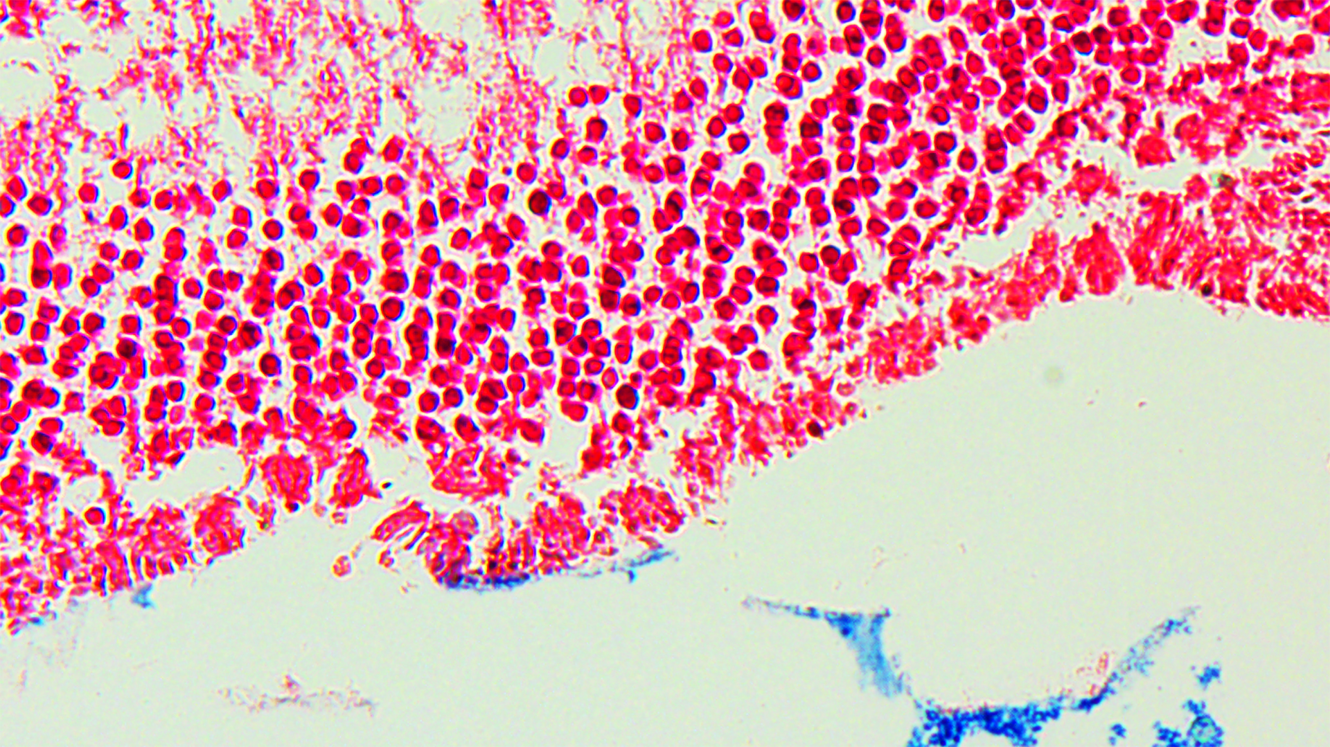
Xenium spatial transcriptomics linked to nuclear morphometry shows that nuclear atypia correlates with rising oncogenic markers and falling homeostatic markers. Fe-NTA exposure generates three karyomegalic niches — quiescent (K1), adaptive (K4), and precancerous (K2) — reflecting distinct nuclear-to-transcriptional states.
Credit: Kong et al., Redox Biology 95 (2026) 104293, CC BY-NC-ND 4.0
Researchers
Related organizations
Related tags
- BRCA1
- Cancer marker
- Cancerous tumors
- Carcinogenesis
- Ferroptosis
- Giant-nucleus cells
- Iron
- Medicine, Dentistry, and Pharmacy
- Oxidative stress
- Pathology