This discovery may pave the way for developing new medications to treat neurodevelopmental disorders.
A Japanese research team has successfully reproduced the human neural circuit in vitro using multi-region miniature organs known as assembloids, which are derived from induced pluripotent stem (iPS) cells. With this circuit, the team demonstrated that the thalamus plays a crucial role in shaping cell type-specific neural circuits in the human cerebral cortex.
These findings were published in the journal Proceedings of the National Academy of Sciences of the United States of America.
Our brain’s cerebral cortex contains various types of neurons, and effective communication among these neurons and other brain regions is crucial for activating functions like perception and cognition.
Patients with neurodevelopmental disorders, such as autism spectrum disorder (ASD), exhibit disruptions in the structure and function of neural circuits in the cerebral cortex. Therefore, understanding the principles of these circuits is essential to uncovering the causes of these disorders and developing new medications.
Previous studies in rodents have demonstrated that the thalamus plays a critical role in shaping neural circuits in the cortex. However, it remains unclear how the interaction between the thalamus and the cortex shapes neural circuit formation in the human cortex.
This uncertainty arises from the ethical and technical challenges involved in collecting brain samples from humans. Organoids are three-dimensional structures that mimic organs and are derived from stem cells. They have gained attention as a novel approach to addressing these kinds of limitations.
However, studying complex biological interactions, such as neural circuit formation, requires more than a single organoid. Assembloids, which are created by combining two or more organoids, allow researchers to effectively replicate parts of the human brain’s neural circuits in vitro.
Professor Fumitaka Osakada, graduate student Masatoshi Nishimura, and their colleagues at the Graduate School of Pharmaceutical Sciences, Nagoya University, established assembloids to reproduce the interaction between the thalamus and the cortex in vitro.
The researchers began by generating cortical and thalamic organoids from human iPS cells. They then physically fused these organoids to create assembloids and studied the thalamus-cortex interactions within them.
Their observations revealed that axons in the thalamus extended toward the cortex, whereas those in the cortex extended toward the thalamus, forming synapses with one another. These findings demonstrated that thalamus-cortex interactions in the assembloid operate similarly to those in the human brain.
They next compared gene expression patterns between the cortical side of the assembloid and a single cortical organoid. They found that the former was more mature than the latter, suggesting that thalamus-cortex interactions promote cortical growth and maturation.
The researchers also examined how these thalamus-cortex interactions contribute to the formation of neural circuits in the cortex of the assembloid. They discovered that neural activity propagates from the thalamus to the cortex in a wave-like pattern, forming a synchronous network within the cortex.
Furthermore, the team measured neural activity levels in three primary subtypes of cortical excitatory neurons: intratelencephalic (IT), pyramidal tract (PT), and corticothalamic (CT). They aimed to identify which types of neurons contribute to synchronous neural activity networks.
The results revealed synchronous activity in PT and CT neurons, both of which project to the thalamus. In contrast, no synchrony was observed in IT neurons. These findings suggest that thalamic inputs selectively promote the formation of cell type-specific synchronized networks, enhancing their functional maturation in the assembloid.
The research team successfully reproduced human neural circuits using assembloids. This achievement provides a system to analyze the origins, structures, and functions of these circuits at the cell-type level.
Osakada concluded, “We have made significant progress in the constructivist approach to understanding the human brain by reproducing it. We believe these findings will help accelerate the discovery of mechanisms underlying neurological and psychiatric disorders, as well as the development of new therapies.”
Paper information:
Masatoshi Nishimura, Shota Adachi, Tomoki Kodera, Akinori Y. Sato, Ryosuke F. Takeuchi, and Fumitaka Osakada. (2025) Thalamus-cortex interactions drive cell type-specific cortical development in human pluripotent stem cell-derived assembloids. Proceedings of the National Academy of Sciences of the United States of America. 122 (47) e2506573122. DOI: https://doi.org/10.1073/pnas.2506573122
Funding information:
This work was supported by the Grants-in-Aid from the Japan Society for the Promotion of Science (F.O.; 20K21476, 21H05168, 22H02771, and 25K02553), PRESTO and CREST from the Japan Science and Technology Agency (F.O.; JPMJPR14F6 and JPMJCR1851), CREST and Brain/MINDS 2.0 from Japan Agency for Medical Research and Development (F.O.; JP23gm1510011 and JP24wm0625110), Hoansha Foundation (F.O.), and SRF Foundation (F.O.).
Expert contact:
Fumitaka Osakada
Graduate School of Pharmaceutical Sciences, Nagoya University
Email: osakada.fumitaka.x3@f.mail.nagoya-u.ac.jp
Media contact:
Naomi Inoue
International Communications Office, Nagoya University
Email: icomm_research@t.mail.nagoya-u.ac.jp
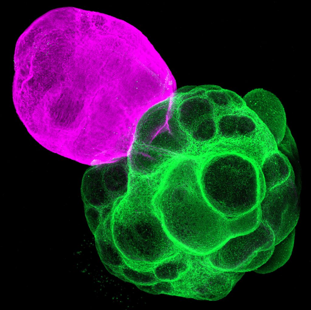
Top image:
Assembloid [3D fluorescent staining]
Axons in the thalamus (pink) extended toward the cortex, while those in the cortex (green) extended toward the thalamus at 14 days post-fusion.
(Credit: Fumitaka Osakada)