Study tracks adult patients before and after rituximab treatment, using rare blood samples from hard-to-treat patients dependent on steroids for a decade.
Nephrotic syndrome is caused when the filtering system in the kidneys is damaged, allowing large amounts of protein to leak into the urine. It is usually treated with steroids, but in some cases steroids alone are insufficient, and long-term steroid use has serious side effects. Some patients develop a difficult-to-treat form of the disease which returns when the steroid dose is reduced.
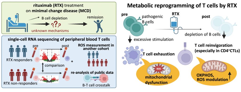
For these patients, the intravenous drug rituximab (RTX) has been found to be effective. RTX removes B cells from the blood, a type of immune cell thought to contribute to the disease. Now, researchers from Nagoya University in Japan have found that the drug does more than eliminate B cells. It also triggers a reaction in another immune cell type, T cells, increasing their energy production and lowering cell stress in patients who respond to RTX treatment. These changes were largely absent in non-responders.
The findings, published in iScience, identify a previously unknown link between B cells and T cells that could help predict how a patient with nephrotic syndrome will respond to RTX.
One drug, two cell types, different responses
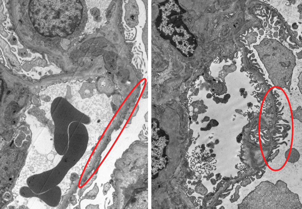
The most common cause of nephrotic syndrome is minimal change disease (MCD). In most cases, it is thought to occur when the immune system damages specialized filtering cells in the kidney, called podocytes. However, the exact mechanism is unknown. Even in severe cases, the kidney shows no visible structural damage under standard examination. The damage only becomes visible under high magnification, hence the name “minimal change.”
“B cells and T cells communicate with each other to regulate the immune system. Normally B cells help fight infections by producing antibodies, but in minimal change disease they are thought to play a role in causing it,” said lead author Eri Koshi-Ito, assistant professor at the Graduate School of Medicine, Nagoya University.
“We tracked changes in gene expression in individual T cells and conducted oxidative stress measurements for these cells in a separate group of patients. All individuals were adults with MCD who had been steroid-dependent for about ten years.”
Blood samples were collected from patients before the first RTX treatment and one month after. Samples from 14 patients were obtained: six for gene expression analysis and eight for oxidative stress measurements. Patients were classified into “responders”, who were able to stop steroids and stay in remission after treatment, and “non-responders”, who could not stop steroids or relapsed within six months of treatment.
Gene expression analysis of T cells showed that genes involved in mitochondrial energy production were more active in responders after treatment. The other patient group showed elevated levels of reactive oxygen species (ROS) in these cells before treatment that dropped after, while non-responders showed the opposite.
Oxidative stress happens when a cell has too many ROS. These are unstable molecules produced as a byproduct of the cell’s energy-making processes. In small amounts, ROS helps the cell function normally, but when they build up faster than the cell can neutralize them, they can damage cell proteins, DNA, and other components, and wear down the cell.
“In both responders and non-responders, RTX depleted B cells as expected, but only in responders did this trigger significant downstream changes in T cells, including a reduction in the number of exhausted T cells weakened by prolonged stimulation. In non-responders, T cells showed comparatively little change,” said senior author Professor Hiroshi Suzuki from the Graduate School of Medicine. “Our findings suggest that the effectiveness of RTX is linked to improved cell metabolism and reduced cell stress soon after treatment begins.”
Using public data on children with nephrotic syndrome, the researchers also found that T cells rely on a less efficient way of producing energy, and that disease-related B cells and T cells show abnormal communication with each other.
Towards a predictive test
B cells nearly disappear after RTX treatment and typically return within six months. Some patients stay in remission well after they return, while others relapse when there are no B cells present. Therefore, B-cell removal alone cannot explain how RTX works, and no reliable method exists to predict who will respond.
In Japan, RTX has only been covered by health insurance for adults since June 2026. For this study, RTX was administered to patients at Nagoya University between 2018 and 2022 under an approved ethical protocol for off-label use. At the time the drug was used more cautiously, often months apart, making it possible to clearly distinguish responders from non-responders. Now that early, repeated RTX use is more common, this distinction is harder to make.
With adults now gaining wider access, who should receive RTX and when remains a key question. Early identification of likely responders could lead to earlier remission and fewer complications. It could also avoid unnecessary repeat infusions and the associated infection risks for likely non-responders.
The researchers believe the early changes they have identified in T-cell metabolism and ROS levels can be used as biomarkers to address these issues and help optimize treatment for this difficult-to-treat form of nephrotic syndrome. They point out that larger trials are needed to confirm their results and determine if these changes can predict how a patient will respond.
Publication information:
Eri Koshi-Ito, Yu Watanabe, Chikao Onogi, Asuka Horinouchi, Koichi Ogami, Seiko Yoshino, Yohei Sugimoto, Shintaro Komatsu, Akihito Tanaka, Kazuhiro Furuhashi, Shoichi Maruyama, and Hiroshi I. Suzuki, 2026. Single-cell transcriptomic signatures of T cells associated with rituximab responsiveness in Idiopathic Nephrotic Syndrome, iScience, 29(9): 117237. DOI: https://doi.org/10.1016/j.isci.2026.117237
Funding information:
This work was supported by grants from Japan Agency for Medical Research and Development (AMED) (JP24ek0109753, JP22ama221111, JP23kk0305026, JP23tk0124003, JP24ck0106875, JP25nk0101727, JP25ck0106019, JP25ak0101291, JP23ck0106791, and JP25kk0305028); YOKOYAMA Foundation for Clinical Pharmacology (YRY-2412); Aichi Kidney Foundation Grant; JSPS Grant-in-Aid for Research Activity Start-up (JP24K23431); JSPS Grant-in-Aid for Early-Career Scientists (JP25K19485, JP25K19486); JSPS Grant-in-Aid for Scientific Research (A) (JP24H00614); JSPS Home-Returning Researcher Development Research (JP19K24694); the Takeda Science Foundation; Toray Science Foundation (22-6304); Inamori Research Institute for Science (InaRIS); and JSPS fellowship for Young Scientists.
Expert contact:
Eri Koshi-Ito
Graduate School of Medicine
Nagoya University
E-mail: ito.eri.r5@f.mail.nagoya-u.ac.jp
Media contact:
Merle Naidoo
International Communications Office
Nagoya University
Email: icomm_research@t.mail.nagoya-u.ac.jp
Top image:
Healthy versus damaged podocytes: An electron microscopy image shows the difference in a healthy kidney (right), with filtering cells called podocytes that have distinct, finger-like structures. In minimal change disease (left), these structures flatten out. This change is only visible at very high magnification. Credit: Eri Koshi-Ito, Nagoya University
Researchers
-
KOSHI EriGraduate School of Medicine, Center for Research of Laboratory Animals and Medical Research Engineering, Division for Advanced Medical Research
-
WATANABE YuGraduate School of Medicine, Center for Research of Laboratory Animals and Medical Research Engineering, Division for Advanced Medical Research
-
OGAMI KoichiGraduate School of Medicine, Center for Neurological Diseases and Cance, Division
-
YOSHINO SeikoGraduate School of Medicine, Center for Research of Laboratory Animals and Medical Research Engineering, Division for Advanced Medical Research
-
TANAKA AkihitoNagoya University Hospital, Cardiology
-
FURUHASHI KazuhiroNagoya University Hospital, Nephrology
-
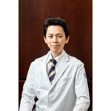 MARUYAMA ShoichiGraduate School of Medicine, Program in Integrated Medicine, Internal Medicine
 -
SUZUKI HiroshiGraduate School of Medicine, Center for Neurological Diseases and Cance, Division