Researchers in Japan revealed that age-related clonal hematopoiesis accelerates aneurysm progression and showed that commonly used osteoporosis drugs could slow or halt this process.

Aortic aneurysms are characterized by abnormal enlargement of the aorta, the primary artery responsible for carrying blood from the heart. Rupture often leads to sudden death, and currently, no effective drug therapies are available to halt disease progression.
Researchers at Nagoya University in Japan found that aortic aneurysms are associated with clonal hematopoiesis, an age-related process in which blood-forming stem cells acquire genetic mutations. Their findings, published in the Journal of Clinical Investigation, suggest that commonly used osteoporosis drugs could slow or halt aneurysm progression.
Currently, surgery is the only definitive treatment for aortic aneurysms. Surgical decisions are guided by the risk of rupture, which is assessed through imaging of aneurysm diameter, morphological features, and expansion rate.
It remains difficult to predict which patients will experience progressive aneurysm enlargement, highlighting the need for additional indicators to better stratify disease progression risk. Furthermore, developing drugs that slow disease progression is crucial for reducing mortality. Achieving both goals requires a clear understanding of the underlying mechanisms.
To address this challenge, Assistant Professor Yoshimitsu Yura and graduate student Jun Yonekawa of the Nagoya University Graduate School of Medicine, along with their colleagues, conducted a comprehensive study.
The research team hypothesized that macrophages derived from clonal hematopoiesis accelerate the progression of aortic aneurysms. Although clonal hematopoiesis is recognized as a contributor to several age-related diseases, such as cardiovascular diseases and osteoporosis, its association with aortic aneurysms remains unclear.
Analysis of patient data
Researchers first conducted a clinical study to examine the relationship between clonal hematopoiesis and abdominal aortic aneurysms in 44 patients scheduled for aneurysm surgery.
Genetic analysis and retrospective clinical data showed that approximately 60% of patients had clonal hematopoiesis. These patients had a significantly faster aneurysm expansion rate compared to those without clonal hematopoiesis.
These results suggest that clonal hematopoiesis, which is detectable through routine blood sampling, may serve as a novel biological marker alongside conventional indicators.
Investigation of causal mechanisms in animal models
Researchers then used a mouse model of clonal hematopoiesis driven by Tet2 mutations. These mice exhibited more rapid aneurysm progression and greater increases in aortic diameter than control mice.
Histological analysis showed thinning and fragmentation of elastin fibers in the aortic wall, substantial macrophage infiltration, and degeneration of adjacent vascular smooth muscle cells.
Further analyses suggested that Tet2-mutant macrophages in affected mice exhibited increased expression of osteoclast-related markers, including TRAP. In vitro, these macrophages showed an enhanced propensity to differentiate into osteoclast-like cells and upregulated MMP-9 expression. These findings suggest a potential mechanism by which Tet2-mutant macrophages may contribute to extracellular matrix degradation and aneurysm progression.
The study also identified the RANK/RANKL signaling axis as a key driver of cellular differentiation. This axis is also involved in the pathogenesis of osteoporosis. Researchers found that inactivating the RANK gene in macrophages suppressed cellular transformation and abnormal aortic expansion.
Potential non-surgical approach
To assess clinical relevance, researchers treated affected mice with osteoporosis drugs—anti-RANKL antibodies and alendronate. This intervention significantly reduced aneurysm progression.
“These drugs could potentially be repurposed for clinical use, as they are already FDA-approved and have established safety profiles,” said Yonekawa, the study’s first author. “Our findings provide a rationale for exploring drug-based therapeutic strategies for aortic aneurysms.”
Yura, the study’s corresponding author, concluded: “Our hypothesis that vascular diseases may result from blood aging enabled us to identify a mechanism underlying aortic aneurysms. We hope these results will improve the prediction of the disease and support the development of treatments to halt progression.”
Paper information:
Jun Yonekawa, Yoshimitsu Yura, Junmiao Luo, Katsuhiro Kato, Shuta Ikeda, Yohei Kawai, Tomoki Hattori, Ryotaro Okamoto, Mari Kizuki, Emiri Miura-Yura, Keita Horitani, Kyung-Duk Min, Takuo Emoto, Hiroshi Banno, Mikito Takefuji, Kenneth Walsh, Toyoaki Murohara (2026). Tet2-driven clonal hematopoiesis drives aortic aneurysm via macrophage-to-osteoclast-like differentiation, The Journal of Clinical Investigation.
DOI: https://doi.org/10.1172/JCI198708
Expert contact:
Yoshimitsu Yura
Nagoya University Graduate School of Medicine
Email: yura.yoshimitsu.z6@f.mail.nagoya-u.ac.jp
Medica contact:
Naomi Inoue
Nagoya University International Communications Office
Email: icomm_research@t.mail.nagoya-u.ac.jp
Top image:
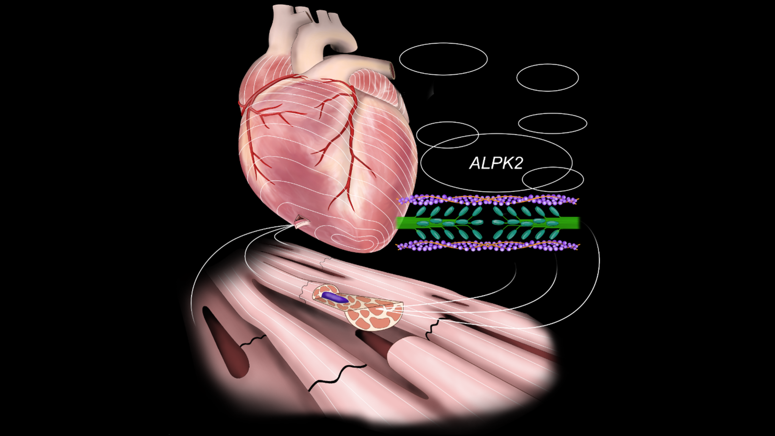
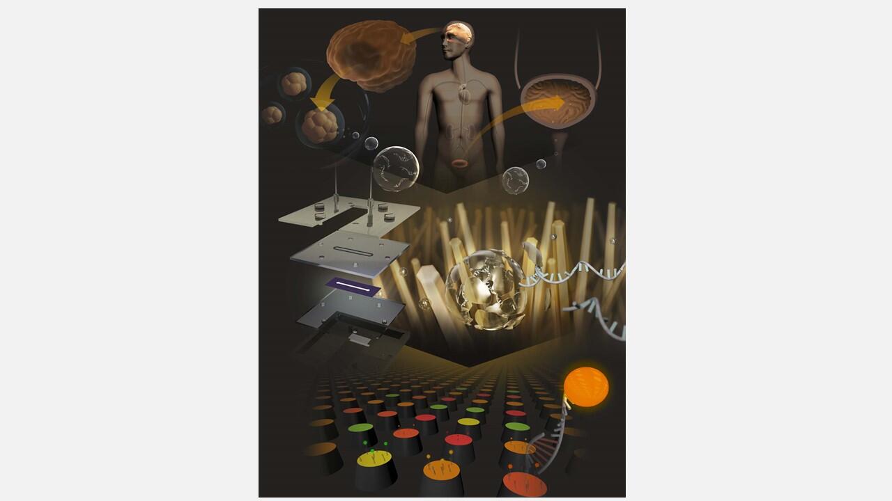
Graphical abstract of the study showing that Tet2-driven clonal hematopoiesis promotes aortic aneurysm progression through macrophage-to-osteoclast-like differentiation.
(Credit: Nagoya University / Jun Yonekawa and Yoshimitsu Yura)