A gene-encoded blueprint tells growing neurons which brain regions to connect with
How complex neural circuits are genetically designed and wired is a fundamental question in neuroscience. Scientists have shown for the first time that genes encode a “wiring map” that guides neurons to connect with the correct brain regions. The findings, based on machine learning analysis of mouse brain data, were published in Proceedings of the National Academy of Sciences, and offer new avenues for research into brain development and disease.
Mapping connections between brain regions with data
The research team, led by scientists from Nagoya University in Japan, aimed to understand the wiring rules that guide nerve fibers during brain development. These long, thin fibers, called axons, extend from neurons and send signals to other neurons.
The researchers developed an analysis method called SPERRFY that combines two datasets. One dataset maps which brain regions are connected to each other, and the other tracks the activity levels of 763 genes in all 213 brain regions in mice.
“Some genes are highly active in certain brain regions and less active in others. These differences create distinct patterns of gene activity throughout the brain,” said Naoki Honda, senior author and professor from Nagoya University’s Graduate School of Medicine. “When hundreds of patterns overlap, they give each brain region a unique molecular identity. These identities are what SPERRFY was designed to decode.”
By feeding both datasets into a machine learning algorithm, SPERRFY identified these patterns of gene activity, called gene expression gradients, that predict which brain regions are likely to connect. For each pair of connected brain regions, SPERRFY paired the gene activity profile of the source region (where the nerve fiber originates) with the profile of the target region it connects to.
From these gene expression gradients, the researchers produced a brain wiring map that tells each brain region where it is relative to every other region. Overlapping patterns of gene activity reconstructed the brain’s connection patterns with a prediction-performance score of 0.88 on a 0-to-1 scale, where 1.0 indicates perfect prediction. By comparison, predictions based only on the physical distance between brain regions scored about 0.70.
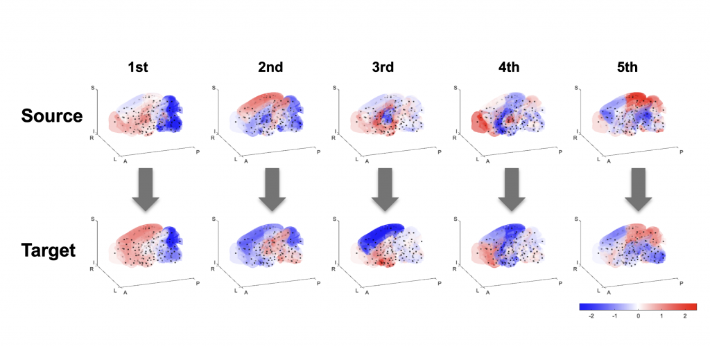
Additionally, the researchers discovered that the brain’s wiring map operates on two levels. Broad gene activity patterns determine the overall organization between brain regions, while more detailed patterns regulate the specific connections within them.
Testing a 60-year-old theory on the whole brain
The findings build on the chemoaffinity theory proposed by Nobel laureate Roger Sperry in 1963. He suggested that neurons find their connection partners by following molecular concentration gradients — chemical signals that vary in strength throughout the brain. These gradients act like a GPS system for growing nerve fibers.
“The chemoaffinity theory was well established for simple circuits such as the visual and olfactory systems. But until now, the complexity of whole-brain connectivity made it difficult to test whether the same principle operates across the brain,” said Jigen Koike, first author and former PhD student at Hiroshima University, who also conducted research as a special research student at Nagoya University’s Graduate School of Medicine.
This complexity made it extremely difficult to test Sperry’s theory across the entire brain without computational tools. Using machine learning, the researchers developed the tools to do this for the first time. Their findings support the idea that this long-standing principle is not limited to simple sensory circuits, but also helps explain how connections are organized across the whole brain.
Future research
By comparing the activity of 763 genes against the wiring map, SPERRFY also identified specific genes with activity patterns that closely matched, including genes known to guide nerve growth. This supports the validity of the method and provides a starting point for research on the molecular mechanisms of brain wiring.
The researchers note that their method can be applied to any species for which maps of the brain’s neural circuits and gene expression data are available, such as humans, marmosets, and fruit flies. As these datasets expand, the method could help determine if the same molecular wiring principles are shared across species and how they have evolved. SPERRFY could also assist scientists in understanding how disruptions in brain wiring contribute to neurodevelopmental disorders.
Paper information:
Jigen Koike, Ken Nakae, Riichiro Hira, Yuichiro Yada, Naoki Honda, 2026. A data-driven framework linking the connectome to spatial gene expression gradients inspired by chemoaffinity theory. Proceedings of the National Academy of Sciences, 123(10). DOI: https://doi.org/10.1073/pnas.2516572123
Funding information:
This work was supported by JST, the establishment of university fellowships toward the creation of science technology innovation (grant number JPMJFS2129), JST SPRING (grant number JPMJSP2132), JSPS KAKENHI (grant number JP22H05163), Moonshot R&D–MILLENNIA Program (grant number JPMJMS2024-9), Agency for Medical Research and Development (AMED) Multidisciplinary Frontier Brain and Neuroscience Discoveries (Brain/MINDS 2.0) (grant number JP25wm0625322 and JP25wm0625210), and the Cooperative Study Program of Exploratory Research Center on Life and Living Systems (ExCELLS: program number 19–102).
Expert contact:
Honda Naoki
Graduate School of Medicine
Nagoya University
Email: honda.naoki.t1@f.mail.nagoya-u.ac.jp
Media contact:
Merle Naidoo
International Communications Office
Nagoya University
Email: icomm_research@t.mail.nagoya-u.ac.jp
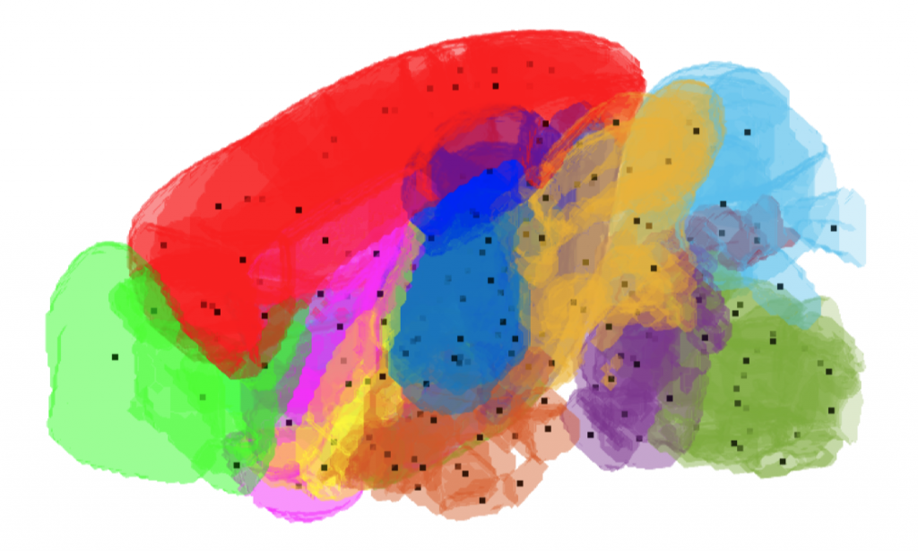
Top image:
A 3D visualization of the 13 major regions in the mouse brain. Black dots mark the centers of the 213 subdivisions used by SPERRFY to analyze relationships between brain connectivity and gene activity patterns. Credit: Koike et al., PNAS, 2026. CC BY 4.0