Study finds floating ovarian cancer cells recruit mesothelial cells in abdominal fluid to create cancer clusters
Ovarian cancer kills more women than any other gynecological cancer. Most patients receive their diagnosis only after the disease spreads throughout the abdomen. Until now, scientists have never fully understood why this cancer advances so fast.
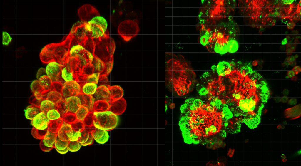
A new study led by Nagoya University explains why. Published in Science Advances, the study shows that cancer cells recruit help from protective mesothelial cells that normally line the abdominal cavity. Mesothelial cells lead the invasion and cancer cells follow the pathways they create. These hybrid cell clusters resist chemotherapy better than cancer alone.
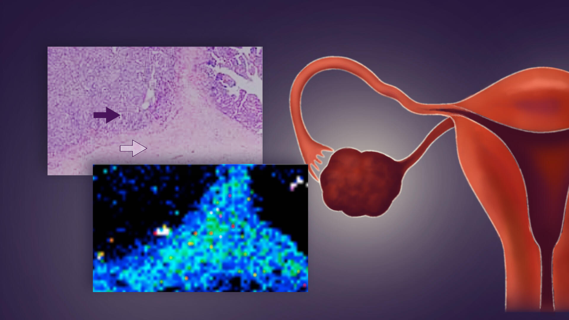
Researchers examined abdominal fluid from ovarian cancer patients and found something unexpected. Cancer cells do not float alone in the abdominal cavity. Instead, they often grab onto mesothelial cells and form hybrid spheres. About 60% of all cancer spheres contain these recruited mesothelial cells. The cancer cells release a protein called TGF-β1 that transforms the mesothelial cells and causes them to develop spike-like structures that cut through tissue.
Invadopodia, spike structures that do the digging for cancer
When ovarian cancer develops, cancer cells break off from the tumor. These cells enter the abdominal fluid and float freely. The fluid moves around as you breathe and move your body. This movement carries the cancer cells to different spots in the abdomen.
Most other cancers spread differently. Breast cancer or lung cancer cells enter blood vessels. They travel through the bloodstream to reach distant organs. Doctors can sometimes track these cancers through blood tests because blood moves in predictable paths through vessels.
Ovarian cancer cells avoid blood vessels entirely. They float in fluid that has no fixed path. This floating stage happens before the cancer cells attach to new organs. Scientists did not fully understand what happened during the floating period or how cells worked together to spread cancer so quickly.
The research team discovered that cancer cells recruit protective mesothelial cells that have shed from the abdominal cavity lining during this floating stage.
The two cell types stick together and form hybrid spheres. The mesothelial cells then grow invadopodia, spike-like structures that drill into surrounding tissue. The hybrid spheres resist chemotherapy drugs more effectively and invade tissues faster when they land on organs.
Outsourcing the hard work of cell invasion
The researchers examined abdominal fluid from ovarian cancer patients using advanced microscopy to watch this process in real time. They confirmed their findings with mouse models and single-cell genetic analysis.
Lead author Dr. Kaname Uno, a former PhD student and current Visiting Researcher at Nagoya University’s Graduate School of Medicine, explained that the cancer cells do not need to become more invasive themselves. “They manipulate mesothelial cells to do the tissue invasion work. They undergo minimal genetic and molecular changes and just migrate through the openings that mesothelial cells create.”
Dr. Uno worked as a gynecologist for eight years before he pursued research. One of his patients changed his career path. She had clear screening results just three months before doctors found advanced ovarian cancer. Current medical tools failed to detect the cancer early enough to save her life. This motivated Dr. Uno to investigate why ovarian cancer spreads so rapidly.
This discovery opens new treatment possibilities. Current chemotherapy targets cancer cells but ignores the mesothelial accomplices. Future drugs could block the TGF-β1 signal or prevent the formation of these dangerous partnerships. The research also suggests that doctors could monitor these cell clusters in abdominal fluid to predict disease progression and treatment response.
Paper Information:
Kaname Uno, Masato Yoshihara, Yoshihiko Yamakita, Kazuhisa Kitami, Shohei Iyoshi, Mai Sugiyama, Yoshihiro Koya, Tomihiro Kanayama, Haruhito Sahara, Satoshi Nomura, Kazumasa Mogi, Emiri Miyamoto, Hiroki Fujimoto, Kosuke Yoshida, Satoshi Tamauchi, Akira Yokoi, Nobuhisa Yoshikawa, Kaoru Niimi, Yukihiro Shiraki9, Jonas Sjölund, Hidenori Oguchi, Kristian Pietras, Atsushi Enomoto, Akihiro Nawa, Hiroyuki Tomita, Hiroaki Kajiyama (2026). Mesothelial cells promote peritoneal invasion and metastasis of ascites-derived ovarian cancer cells through spheroid formation, Science Advances, 21(6). https://doi.org/10.1126/sciadv.adu5944
Funding information:
This study was supported by Japan Society for the Promotion of Science (JSPS) KAKENHI Grants-in Aid for Scientific Research grant numbers 20H03824, 21KK0157, 21KK0296, 21K16788, 23K18326, and 24K12529; JSPS Overseas Research Fellowships (202460603); JST FOREST Program (JPMJFR235L); and Lena Wäppling’s Foundation (2025).
Expert Contact:
Kaname Uno
Graduate School of Medicine
Nagoya University
Email: kaname.uno@med.lu.se
Media contact:
Merle Naidoo
International Communications Office
Nagoya University
Email: icomm_research@t.mail.nagoya-u.ac.jp
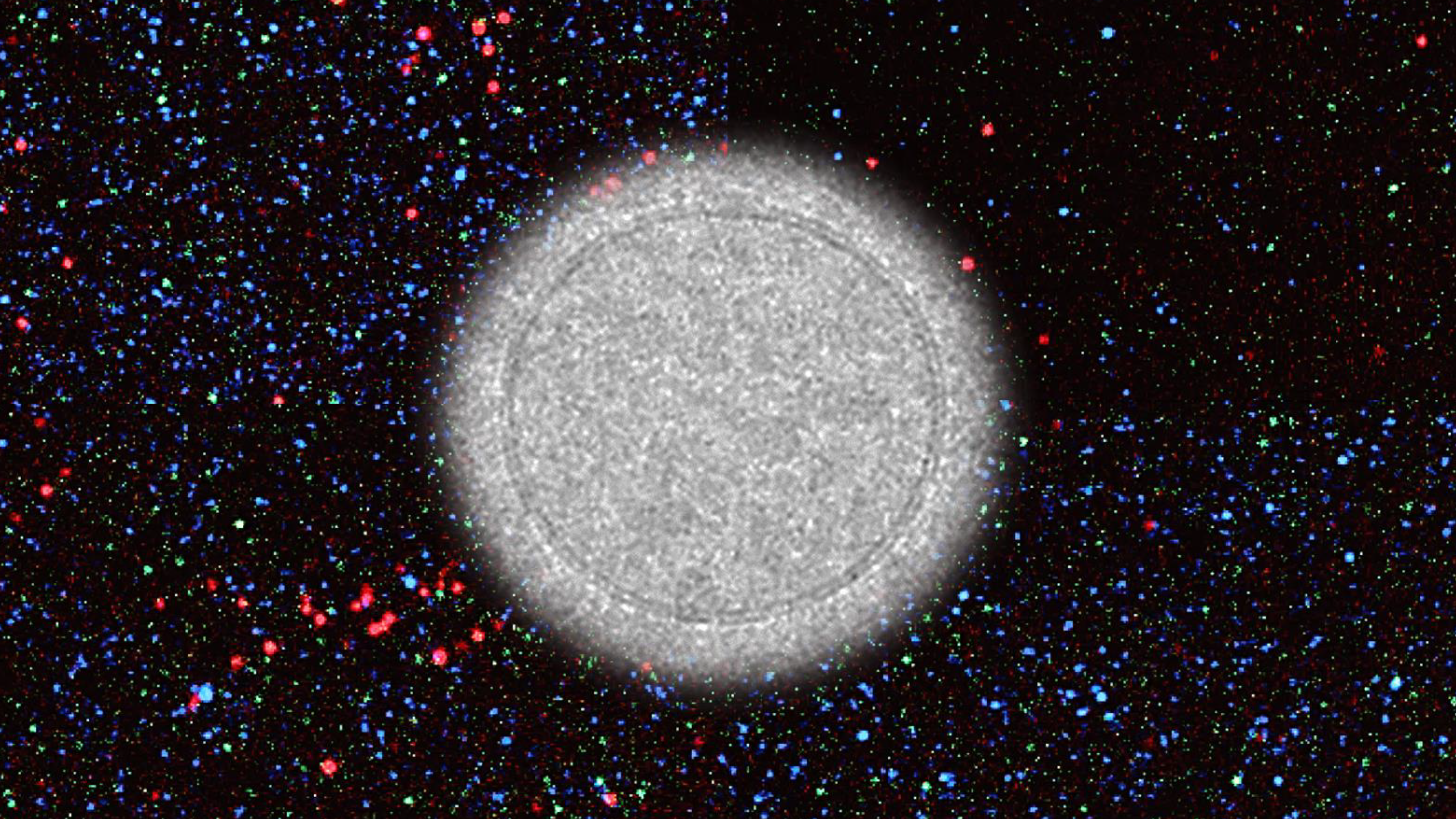
Top image:
Cancer cells (red) stick to mesothelial cells (green) and form hybrid spheres that cut into surrounding abdominal tissue. Credit: Uno et al., 2026
Researchers
-
KAJIYAMA HiroakiGraduate School of Medicine, Program in Integrated Medicine, Medicine in Growth and Aging
-
YOSHIHARA MasatoNagoya University Hospital, Obstetrics and Gynecology
-
IYOSHI ShoheiInstitute for Advanced Research
-
MOGI KazumasaNagoya University Hospital
-
YOSHIDA KosukeNagoya University Hospital, Obstetrics and Gynecology
-
TAMAUCHI SatoshiNagoya University Hospital, Obstetrics and Gynecology
-
YOKOI AkiraNagoya University Hospital, Obstetrics and Gynecology
-
YOSHIKAWA NobuhisaNagoya University Hospital, Obstetrics and Gynecology
-
NIIMI KaoruGraduate School of Medicine, Program in Integrated Medicine, Medicine in Growth and Aging
Related organizations
Related tags
- Cancer research
- Invadopodia
- Medicine, Dentistry, and Pharmacy
- Mesothelial cells
- Ovarian cancer
- Tumor microenvironment