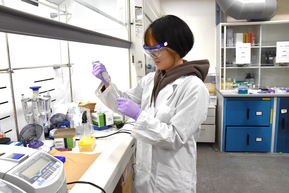
A new technique to diagnose chronic inflammation developed at Nagoya University can read information from the surface and within individual extracellular vesicles in blood and urine. This technique could be used to develop non-invasive tests that can not only detect chronic inflammation but also provide information about the tissues where it occurs.
Whenever our body faces damage from injury, illness, or toxins, it launches an inflammatory response that involves dilating blood vessels to rush fluid and nutrients to the affected site and sending inflammatory cells to destroy pathogens and repair tissue.
But sometimes, the body’s immune system can keep sending inflammatory cells to attack tissues or organs over months or years, even in the absence of any threat. Known as chronic inflammation, it underlies cancers, brain disorders, autoimmune diseases and heart disease, and it gradually causes irreversible tissue damage.
Early detection of chronic inflammation is important because prolonged inflammation can cause irreversible tissue damage and contribute to disease progression.
“Currently, there are several conventional inflammatory biomarkers such as C-reactive protein,” said Taiga Ajiri, an assistant professor at Nagoya University’s Yasui Lab, part of the Department of Biomolecular Engineering. “These biomarkers can detect inflammation, but it is difficult to identify where it occurs.”
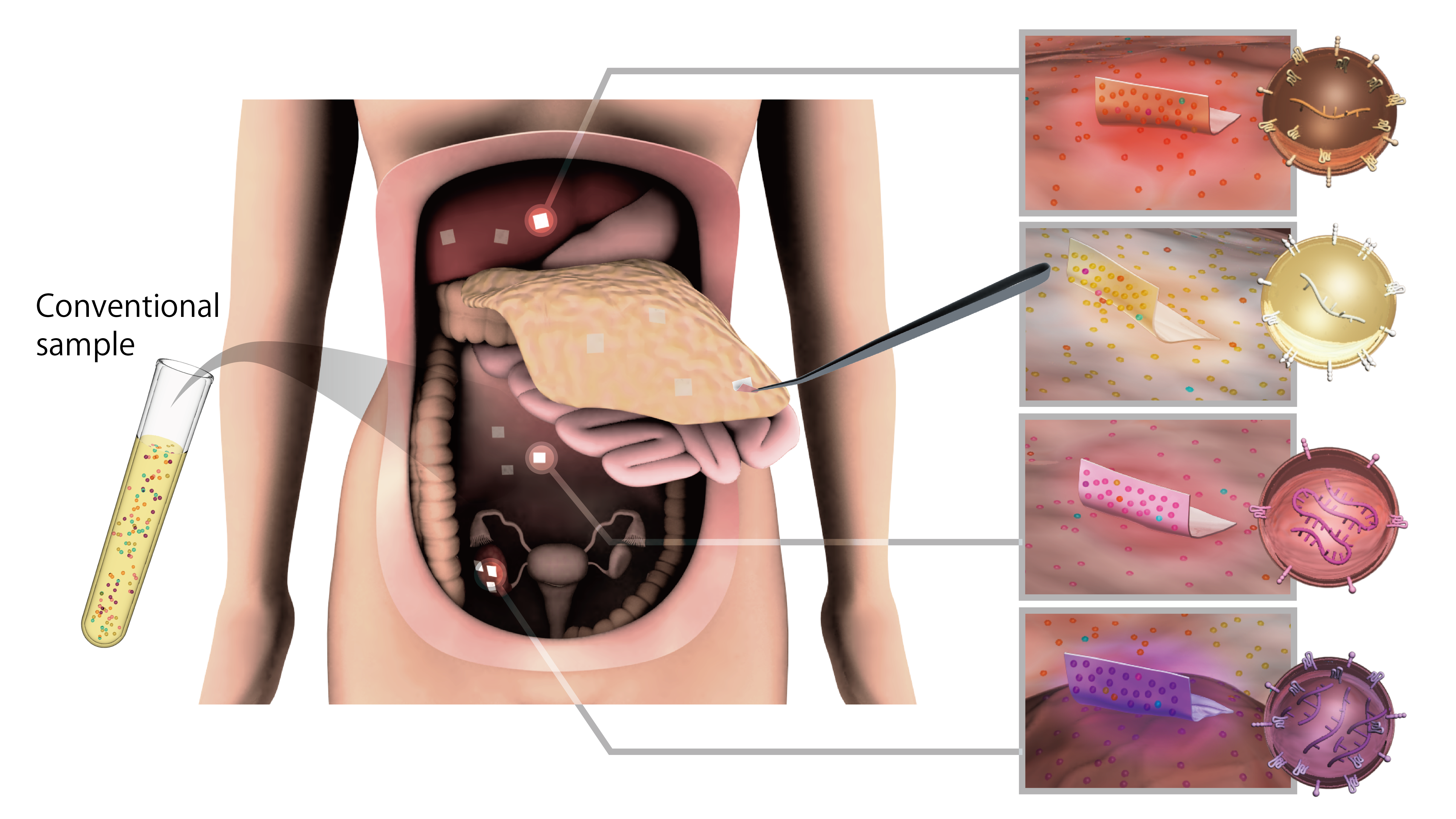
To obtain clues about where inflammation occurs, the researchers turned to extracellular vesicles (EVs): tiny membrane-bound particles released by cells throughout the body that carry molecular information from their cells of origin.
EVs work like parcels with tracking labels. Proteins on their membranes can provide clues about which tissues they came from, while microRNAs inside EVs can reflect the condition of their cells of origin. The vesicle membrane protects this cargo as it travels through body fluids such as blood and urine.
Conventionally, measuring internal microRNAs often requires breaking open EV membranes, mixing molecules from many vesicles together. This makes it difficult to link information inside a vesicle to proteins on its surface.
But reading both internal microRNA and surface protein information without breaking the EV membrane could reveal previously hidden EV subpopulations.
Reading an EV package
The researchers used cellulose nanofiber (CNF) sheets made from wood-derived cellulose. Their mesh-like nanoscale pores can capture and retain EVs. Previous work by Professor Takao Yasui’s group established the use of these sheets for EV capture and analysis.
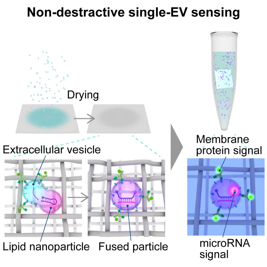
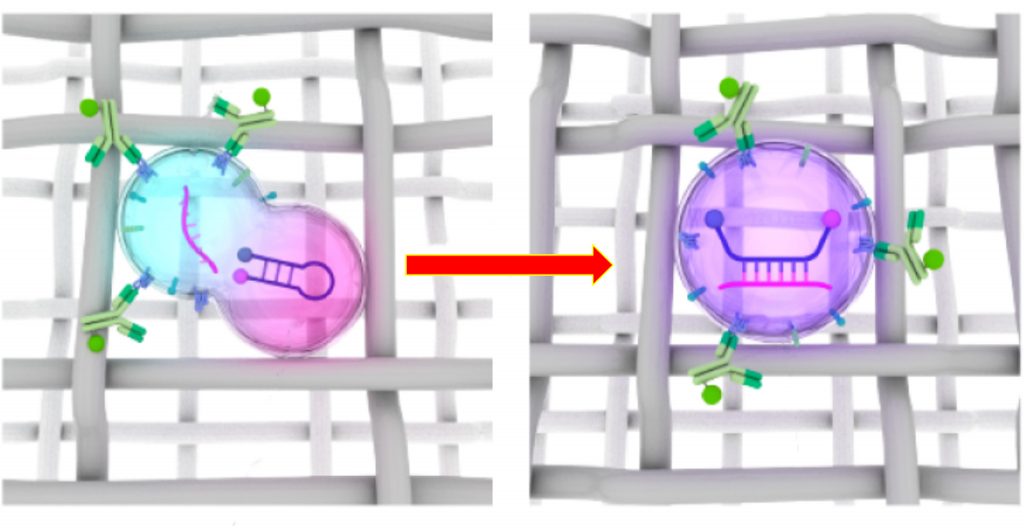
The team found that the drying process on CNF sheets can bring membrane-bound particles into close contact and promote membrane fusion. They applied this property to combine EVs with lipid nanoparticles containing molecular beacons, which are probes that detect specific microRNAs.
“These lipid nanoparticles can deliver molecular beacons inside the EV without disrupting the EV,” Ajiri said. These molecular beacons emit fluorescence when they bind to specific microRNAs, allowing researchers to detect the presence of these target molecules.
Clues to the source: kidney inflammation
To test the method, the team established a laboratory model of chronic active antibody-mediated rejection (CAAMR), a complication of kidney transplantation, by exposing human kidney cells to inflammatory signals. They identified 71 microRNAs with altered expression in released EVs and focused on the highly abundant miR-1246. They also selected DPYS, a kidney-associated membrane protein, to combine information on inflammation with information on tissue origin.
After accounting for detection efficiency, the researchers estimated that about one in ten EVs from unstimulated kidney cells contained miR-1246, compared with about four in ten after inflammatory stimulation. By contrast, qPCR analysis using equivalent sample inputs did not detect a statistically significant difference.
The team then analyzed urine from kidney transplant patients with CAAMR, patients with immune diseases not primarily involving the kidney, and healthy volunteers.
Neither DPYS nor miR-1246 alone clearly distinguished the patient groups. EVs carrying both miR-1246 and the general EV marker CD63 were increased in inflammatory disease groups, but this combination could not distinguish renal from non-renal inflammation. In contrast, EVs carrying both DPYS and miR-1246 were enriched in CAAMR patients, providing clues to inflammation associated with the transplanted kidney.
The team hopes to improve particle recovery and streamline the CNF-based procedure while testing the approach in larger clinical studies. Because urine and blood can be collected with relatively little burden, the method may contribute to less-invasive evaluation and monitoring of chronic inflammatory diseases. These findings have been published in the journal Cell Reports Physical Science.
Publication information:
Shota Kawaguchi, Taiga Ajiri, Kunanon Chattrairat, Hirotaka Koga, Yukiko Kamiya, Fuminori Sato, Hiroyuki Asanuma, Masatoshi Maeki, Manabu Tokeshi, Takeshi Yokoyama, Yoshikazu Tanaka, Kosuke Yoshida, Akira Yokoi, Kiyohiko Hotta, Naoya Iwahara, Yuichiro Fujieda, Mitsutaka Yasuda, Tatsuya Atsumi, Shimpei I. Kubota, Kaoru Murakami, Masaaki Murakami, Takao Yasui, 2026. Non-destructive colocalization sensing of microRNAs and membrane proteins in single vesicles reveals spatial inflammation signatures. Cell Reports Physical Science. DOI:10.1016/j.xcrp.2026.103613
Funding information:
This research was supported by the Moonshot Research and Development Program (grant nos. 22zf0127004s0902 and JP22zf0127009) from the Japan Agency for Medical Research and Development (AMED) (to T. Yasui), AMED (JP243fa627005, JP21zf0127004, and JP23gm1910005 to M. Murakami; 24ae0121056 to H.A.), the New Energy and Industrial Technology Development Organization (NEDO) JPNP20004 (to T. Yasui and H.K.), Q-Leap (JPMXS0120330644 to M. Murakami), the Japan Science and Technology Agency (JST) CREST (JPMJCR2576 to T. Yasui), AIP Acceleration Research (JPMJCR23U1 to T. Yasui), and FOREST (JPMJFR2003 to H.K.); the JSPS Grant-in-Aid for Scientific Research (A) 24H00792 (to T. Yasui); the JSPS Grant-in-Aid for Scientific Research (S) 21H05025 (to H.A.); the JSPS Grant-in-Aid for Scientific Research (S) 24H00038 (to M.T.); the JSPS Grant-in-Aid for Scientific Research (B) 23K28408 (to M. Maeki); the JSPS Grant-in-Aid for Scientific Research (A) 23H00541 (to M. Maeki); the JSPS Grant-in-Aid for Transformative Research Areas (B) 25H01414 (to Y.K.); JSPS Grant-in-Aid for Young Scientists (Start-up) 24K23101 (to T.A.); Platform Project for Supporting Drug Discovery and Life Science Research (Basis for Supporting Innovative Drug Discovery and Life Science Research [BINDS]) from AMED under grant no. JP24ama121038 (support no. 4008) (to T. Yokoyama); Grant for Joint Research Program of the Institute for Genetic Medicine, Hokkaido University, by the Photo-excitonic Project, Hokkaido University, and by the Promotion Project for Young Investigators at Hokkaido University, Japan (to M. Murakami); Science Tokyo SPRING grant no. KH240019nn (to S.K.); JSPS Research Fellowship 25KJ1250 (to S.K.); and the Noguchi Institute NJ202308 (to T. Yasui).
Expert contacts:
Takao Yasui
Department of Biomolecular Engineering,
Graduate School of Engineering,
Nagoya University
Email: yasui@chembio.nagoya-u.ac.jp
Shota Kawaguchi
Department of Life Science and Technology,
Institute of Science Tokyo
Email: kawaguchi.s.5a42@m.isct.ac.jp
Taiga Ajiri
Department of Biomolecular Engineering,
Graduate School of Engineering,
Nagoya University
Email: ajiri@chembio.nagoya-u.ac.jp
Media contact:
Sumeet Kulkarni
International Communications Office
Nagoya University
Email: icomm_research@t.mail.nagoya-u.ac.jp
Researchers

Related organizations
Related tags
- Biology
- Chronic inflammation
- Extracellular vesicle
- Extracellular vesicles
- inflammation
- Kidney disease
- Kidney Health
- Lipid nanoparticle
- Medicine, Dentistry, and Pharmacy